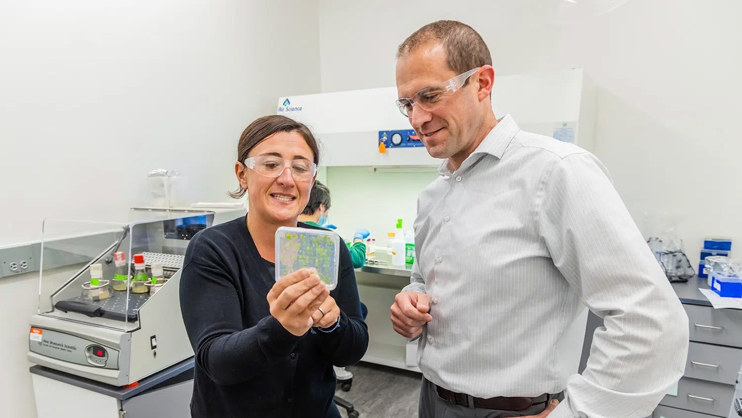
New Technique for Sorting Live Cells May Expedite Biomedical Research
Researchers from North Carolina State University and University of North Carolina at Chapel Hill have developed a new technique that uses sound waves to rapidly separate selected collections of cells for use in biomedical research.
“We think this is important because it will make it faster and easier for researchers to sort out the live cells they need for research ranging from disease study to drug development,” says Dr. Xiaoning Jiang, an associate professor of mechanical and aerospace engineering and adjunct professor of biomedical engineering at NC State and co-author of a paper on the work.

Biomedical research often focuses on how specific cell types respond to various chemicals or environmental factors. These cells are often grown in a liquid medium and on top of a collection of “micropallets,” which are essentially small plastic platforms that sit on the substrate at the bottom of the container. Researchers then select the cells they want and detach the relevant micropallets, which can be removed for additional experimentation or analysis.
Current techniques for removing these micropallets rely on lasers or physical manipulation to separate the pallets from the substrate. But each approach has its drawbacks. Physical manipulation is a slow process, while the energy produced by lasers to release larger micropallets (e.g., a micropallet 500 micrometers in diameter) can inadvertently kill a significant number of the cells. Neither technique is efficient at detaching a significant number of large micropallets quickly.
The new technique from NC State uses ultrasound technology to release the micropallets. Specifically, it uses focused, relatively high-frequency sound waves that are translated into a wave of pressure within the substrate itself. When that wave of force hits a targeted micropallet, the pallet is lifted off the substrate and can be removed, together with its attached cells, for further study. Video of a micropallet being released can be seen here.
Using this technique, micropallets can be selectively released in less than a millisecond. This is not as fast as laser-based techniques, but is much faster than physical manipulation. However, the ultrasound technique has a viability rate of better than 90 percent, meaning that more than 90 percent of live cells survive the process. This is significantly better than existing techniques for the release of large-sized pallets, which can have viability rates of less than 50 percent.
The paper, “Ultrasound-Induced Release of Micropallets with Cells,” was published online Oct. 16 in Applied Physics Letters. Lead author of the paper is Sijia Guo, a Ph.D. student at NC State. Co-authors are Jiang; Dr. Nancy Allbritton, head and professor of the joint Biomedical Engineering Department at NC State and the University of North Carolina at Chapel Hill and professor of chemistry at UNC; and Dr. Yuli Wang, a research associate in the Chemistry Department at UNC-Chapel Hill. The research was supported by a grant from the National Institutes of Health.
-shipman-
Note to Editors: The study abstract follows.
“Ultrasound-Induced Release of Micropallets with Cells”
Authors: Sijia Guo, Nancy Allbritton and Xiaoning Jiang, North Carolina State University; Yuli Wang, University of North Carolina at Chapel Hill
Published: Online Oct. 16 in Applied Physics Letters
Abstract: Separation of selected adherent live cells attached on an array of microelements, termed micropallets, from a mixed population is an important process in biomedical research. We demonstrated that adherent cells can be safely, selectively and rapidly released from the glass substrate together with micropallets using an ultrasound wave. A 3.3-MHz ultrasound transducer was used to release micropallets (500 [micrometers] × 500 [micrometers] × 300 [micrometers]) with attached HeLa cells, and a cell viability of 92% was obtained after ultrasound release. The ultrasound-induced release process was recorded by a high-speed camera, revealing a proximate velocity of ~0.5 m/s.


