Smart Coating Opens Door To Safer Hip, Knee and Dental Implants
Researchers at North Carolina State University have developed a “smart coating” that helps surgical implants bond more closely with bone and ward off infection.
When patients have hip, knee or dental replacement surgery, they run the risk of having their bodies reject the implant. But the smart coating developed at NC State mitigates that risk by fostering bone growth into the implant. The coating creates a crystalline layer next to the implant, and a mostly amorphous outer layer that touches the surrounding bone. The amorphous layer dissolves over time, releasing calcium and phosphate, which encourages bone growth.

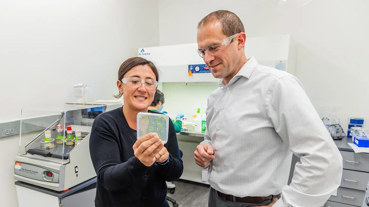
“The bone grows into the coating as the amorphous layer dissolves, resulting in improved bonding, or osseointegration,” says Dr. Afsaneh Rabiei, an NC State associate professor of mechanical and aerospace engineering, associate faculty member of biomedical engineering and co-author of a paper describing the research. This bonding also makes the implant more functional, because the bonding helps ensure that the bone and the implant do a better job of sharing the load.
“We call it a smart coating because we can tailor the rate at which the amorphous layer dissolves to match the bone growth rate of each patient,” Rabiei says. This is important because people have very different rates of bone growth. For example, young people’s bones tend to grow far faster than the bones of older adults.
The researchers have also incorporated silver nanoparticles throughout the coating to ward off infections. Currently, implant patients are subjected to an intense regimen of antibiotics to prevent infection immediately following surgery. However, the site of the implant will always remain vulnerable to infection. But by incorporating silver into the coating, the silver particles will act as antimicrobial agents as the amorphous layer dissolves, Rabiei says. This will not only limit the amount of antibiotics patients will need following surgery, but will provide protection from infection at the implant site for the life of the implant. Moreover, the silver is released more quickly right after surgery, when there is more risk of infection, due to the faster dissolution of the amorphous layer of the coating. Silver release will slow down while the patient is healing. “That is another reason why we call it smart coating,” Rabiei says.
The research was funded by the National Science Foundation, and was accomplished with assistance from the Center for Nanophase Materials Sciences and Shared Research Equipment User Facilities at Oak Ridge National Laboratory.
The research, “Functionally graded hydroxyapatite coatings doped with antibacterial components,” was co-authored by Rabiei, former NC State Ph.D. student Xiao Bai, and Oak Ridge National Laboratory researchers Karren More and Christopher Rouleau. The research is published online by Acta BioMaterialia.
-shipman-
Note to editors: The study abstract follows.
“Functionally graded hydroxyapatite coatings doped with antibacterial components”
Authors: Xiao Bai, Afsaneh Rabiei, North Carolina State University; Karren More, Christopher M. Rouleau, Oak Ridge National Laboratory
Published: Online January 2010, Acta BioMaterialia
Abstract: A series of functionally graded hydroxyapatite (FGHA) coatings incorporated with various percentages of silver were deposited on titanium substrates using ion beam-assisted deposition. The analysis of the coating’s cross-section using transmission electron microscopy (TEM) and scanning transmission electron microscopy equipped with energy dispersive X-ray spectroscopy has shown a decreased crystallinity as well as a distribution of nanoscale (10–50 nm) silver particles from the coating/substrate interface to top surface. Both X-ray diffraction and fast Fourier transforms on high-resolution TEM images revealed the presence of hydroxyapatite within the coatings. The amount of Ag (wt.%) on the outer surface of the FGHA, as determined from X-ray photoelectron spectroscopy, ranged from 1.09 to 6.59, which was about half of the average Ag wt.% incorporated in the entire coating. Average adhesion strengths evaluated by pull-off tests were in the range of 83 ± 6 to 88 ± 3 MPa, which is comparable to 85 MPa for FGHA without silver. Further optical observations of failed areas illustrated that the dominant failure mechanism was epoxy failure, and FGHA coating delamination was not observed.
- Categories: