Forensic Researchers Set Standards for X-Ray Identification of Bodies
For Immediate Release
Forensic researchers have for the first time established science-based standards for identifying human remains based on X-rays of an individual’s spine, upper leg or the side of the skull.
“In the past, forensic experts have relied on a mixed bag of standards when comparing ante mortem and post mortem X-rays to establish a positive identification for a body – but previous research has shown that even experts can have trouble making accurate identifications,” says Ann Ross, lead author of a paper on the new standards and a professor of anthropology at North Carolina State University.
“We’ve created a set of standards that will allow for a consistent approach to identification – that can be replicated – and that allows experts to determine probabilities for an identification,” Ross says. “For example, you could say with 85 percent certainty that a body is a specific individual.”
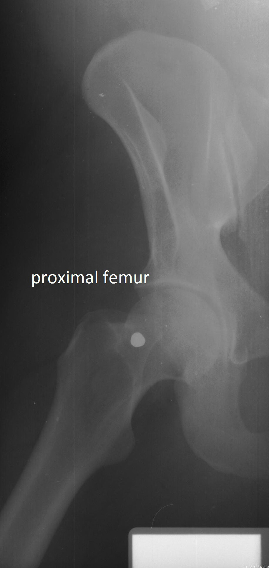
The researchers compared ante mortem and post mortem lateral craniofacial (side of the skull) X-rays for 20 individuals, and did the same for X-rays of the vertebral column (spine) for 50 individuals, and X-rays of the proximal femur (upper leg) for 23 individuals. The researchers used these evaluations to develop location-specific standards for each skeletal region. The researchers focused on these skeletal regions because they are among the most frequently X-rayed in a clinical setting.
The researchers then used additional, unmatched X-rays to test the accuracy of the standards in two ways. First, they tested the standards to see how likely they were to accurately identify a body. Second, they ran separate analyses to see how likely the standards were to “misclassify” an identification – to provide a false-positive or false-negative result.
The standards varied significantly.
For example, a side X-ray of the skull could be used to identify a body with 97 percent certainty and a 10 percent misclassification rate based on as few as two specific traits – as long as there were no inconsistencies in the shapes of the skull X-rays. Cervical – or neck – vertebrae performed even better. These could provide greater than 98 percent certainty and a 7 percent misclassification rate based on a single trait.
At the other end of the spectrum, lumbar – or lower back – vertebrae had a 40 percent misclassification rate even if four matching traits were identified.
“Lumbar X-rays can be used to support information from other skeletal locations, but we think they are too unreliable to be used as the primary means of identification,” Ross says.
In addition to the location-specific standards, the researchers developed a “decision tree” that forensic experts can use to determine the probability of an ID.
“The goal here is to bring standards and consistency to radiographic ID, and to bring some quantification to the process – so that it’s not just based on one expert’s individual opinion,” Ross says.
“We hope to build on these standards by compiling and evaluating a larger database of post mortem and ante mortem X-rays for these and other skeletal structures,” Ross adds.
The paper, “Establishing Standards for Side-by-Side Radiographic Comparisons,” is published online in the American Journal of Forensic Medicine and Pathology. The paper was co-authored by Alicja Lanfear of Middle Tennessee State University and Ashley Maxwell of the University of South Florida. The work was supported by the National Institutes of Justice under grant number 2010-DN-BX-K214.
-shipman-
Note to Editors: The study abstract follows.
“Establishing Standards for Side-by-Side Radiographic Comparisons”
Authors: Ann H. Ross, North Carolina State University; Alicja K. Lanfear, Middle Tennessee State University; Ashley B. Maxwell, University of South Florida
Published: March 17, American Journal of Forensic Medicine and Pathology
DOI: 10.1097/PAF.0000000000000223
Abstract: The objectives of this research were to evaluate the use of various anatomical features that are visible in standard radiographs and to develop a standard system of assessing concordant features for making positive identifications through radiographic comparison. The radiographs used in the study include craniofacial (n = 41), chest (n = 100), and proximal femur (n = 49), which were made available by the North Carolina Office of the Chief Medical Examiner. Radiographs were scored for number of concordant features and were analyzed using classification decision trees. The accuracy of the classification tree models was evaluated using a receiver operating characteristic. Two or more points of concordance are required in lateral cranial radiographs for a 97% probability of a positive identification. If more than 1 concordant feature exists on the cervical vertebrae, there is a 99% probability of correct identification. For thoracic and lumbar vertebrae, 4 or more concordant features are required for a 98% probability of correct identification. If there are 1 or more femoral head and neck concordant features, the probability of a correct identification is 94% and 97%, respectively. This study established the minimum number of concordant areas needed to confirm positive identifications in 3 standard radiographic views.
- Categories:


