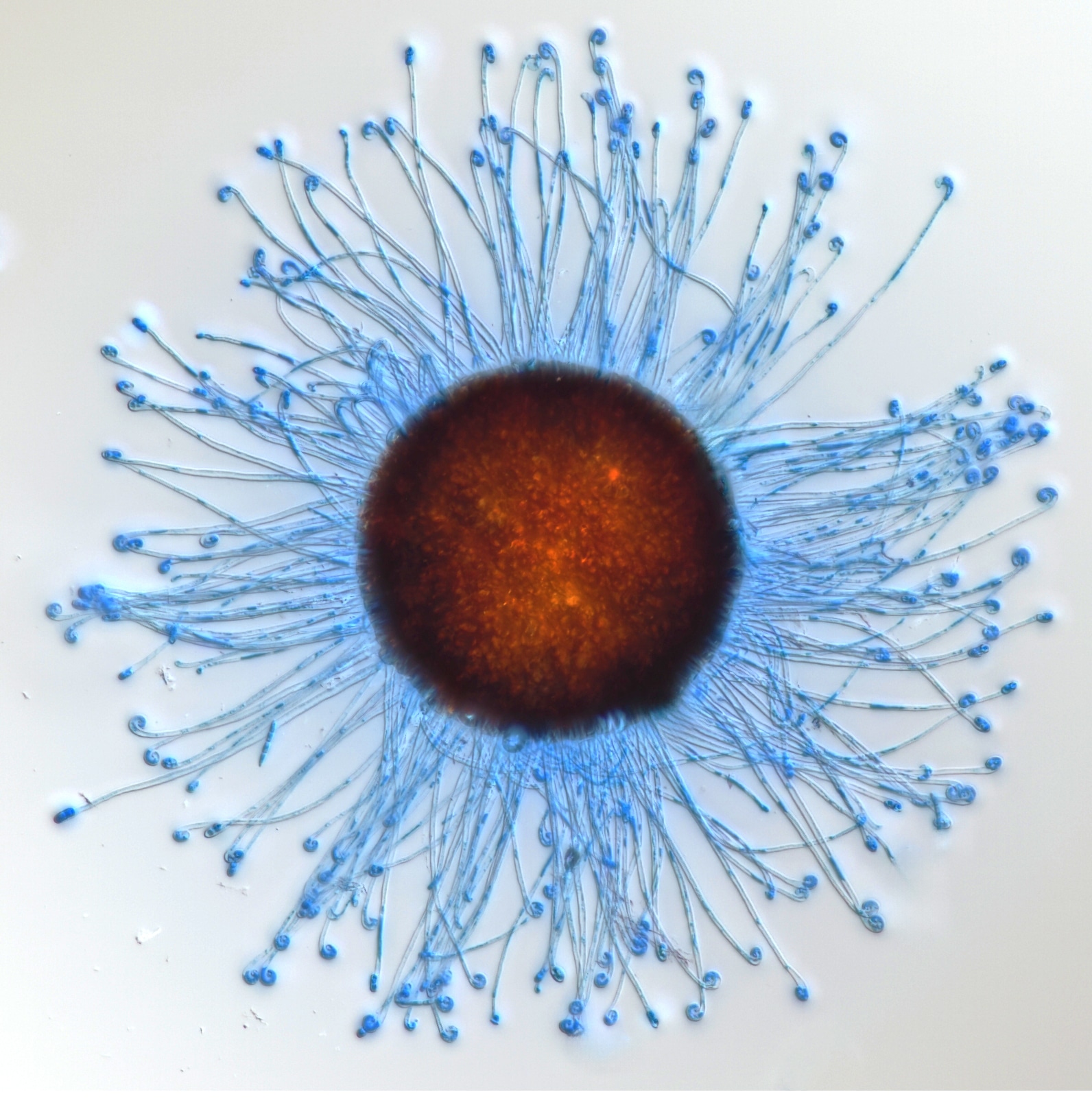
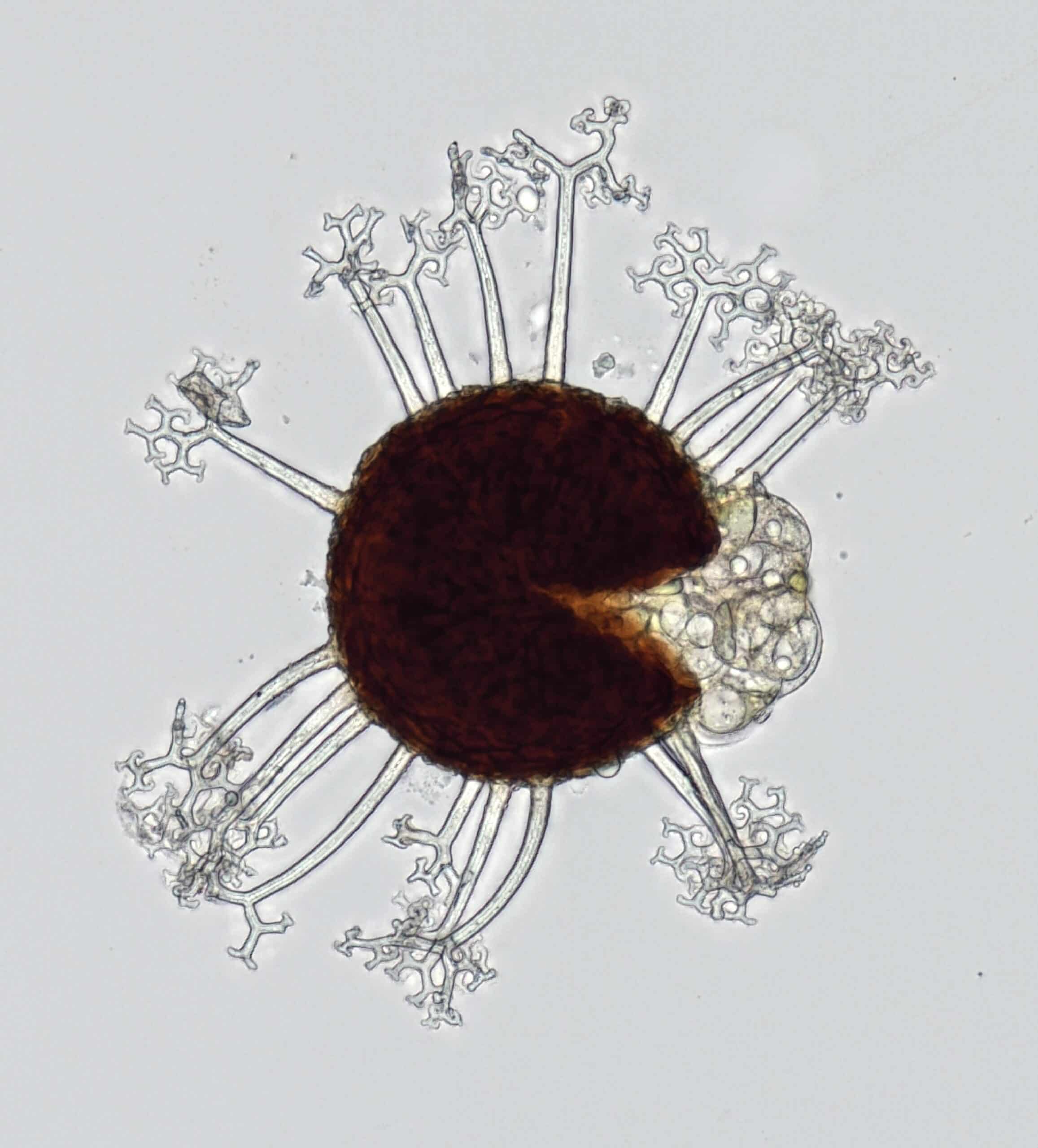
Microscopy: Honorable mention, Undergraduate Students: Christian Shaw for “Takamatsuella circinata chasmothecium dyed with lactophenol blue and imaged using a compound light microscope.”
From volcanic eruptions to microscopic polymers, NC State researchers highlighted their vital work in natural resources, biochemistry, engineering and more through art in the 2024 Envisioning Research Contest. Explore a selection of photos, graphics, data visualization and microscopic images from this year’s contest in the gallery below. A complete list of the winners is here.
The contest is a collaborative effort involving NC State’s Office of Research and Innovation, The Graduate School, the NC State University Libraries, the Office of Undergraduate Research, and University Communications and Marketing. The Envisioning Research contest was open to faculty, staff, graduate students, postdoctoral researchers and undergraduates.
See all the winners, including videos and interactive graphics, on Flickr. Images from the Envisioning Research contest will be exhibited on the Art Wall in the Hunt Library on Centennial Campus.
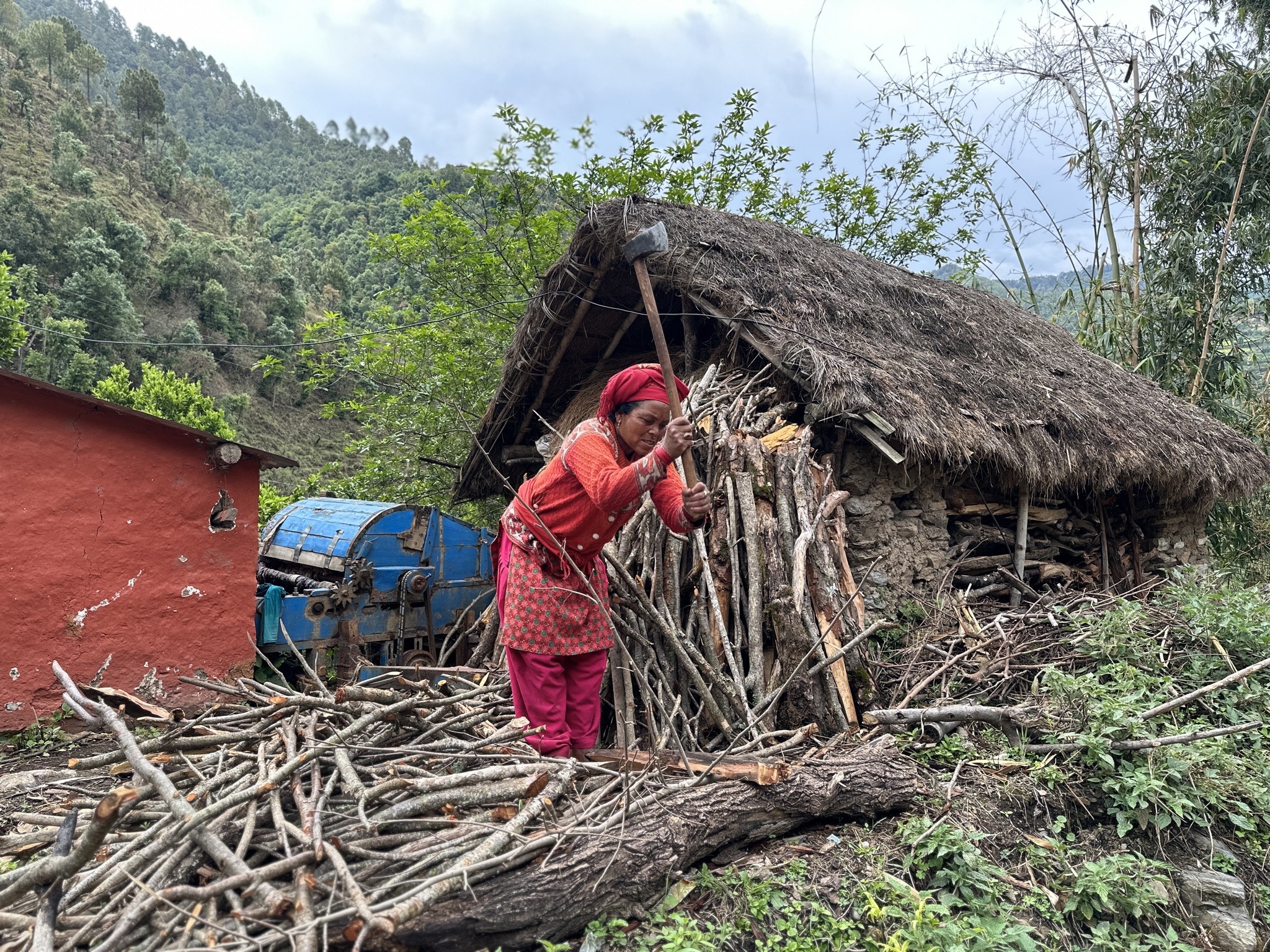
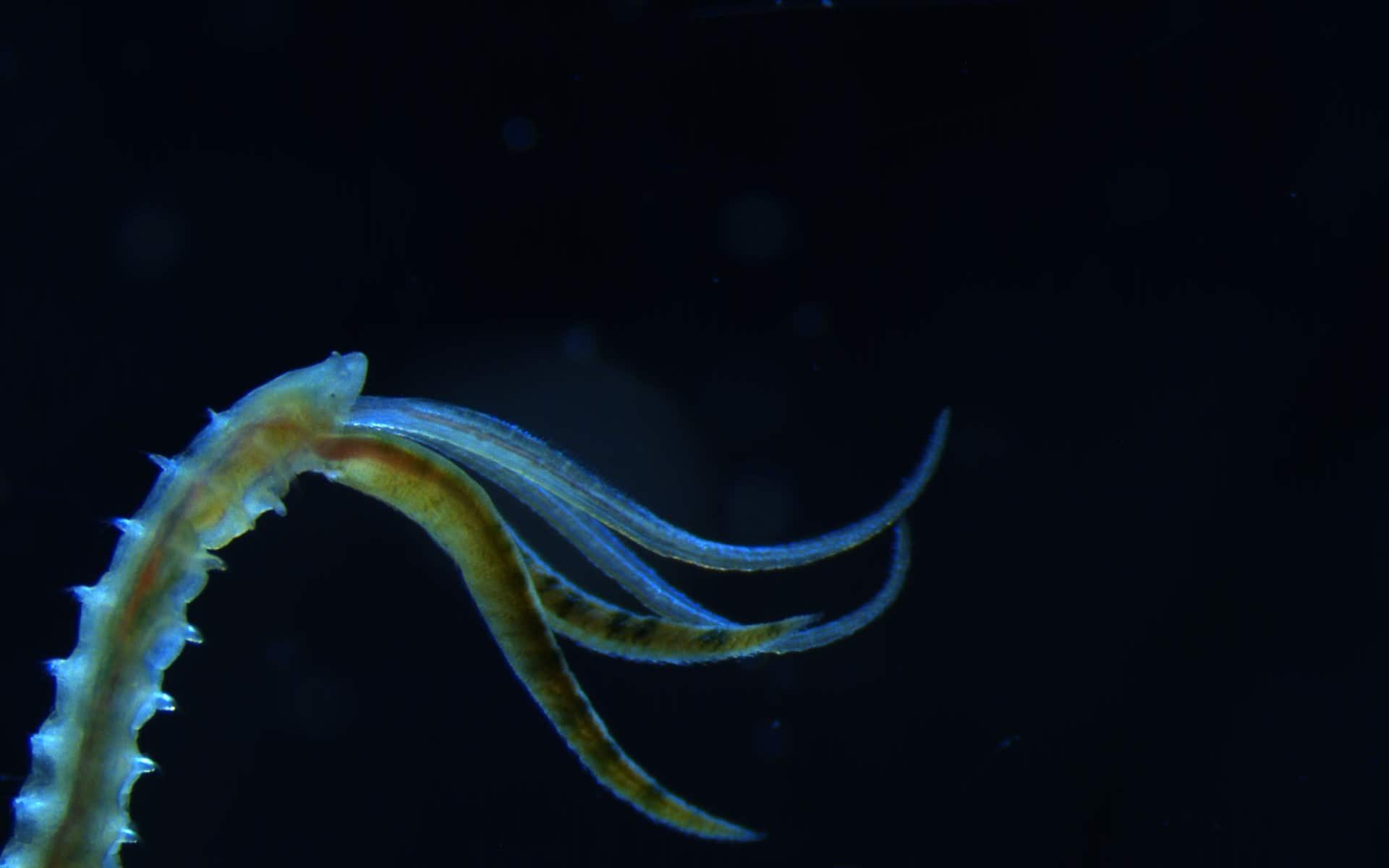
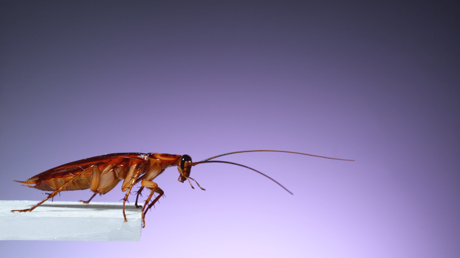
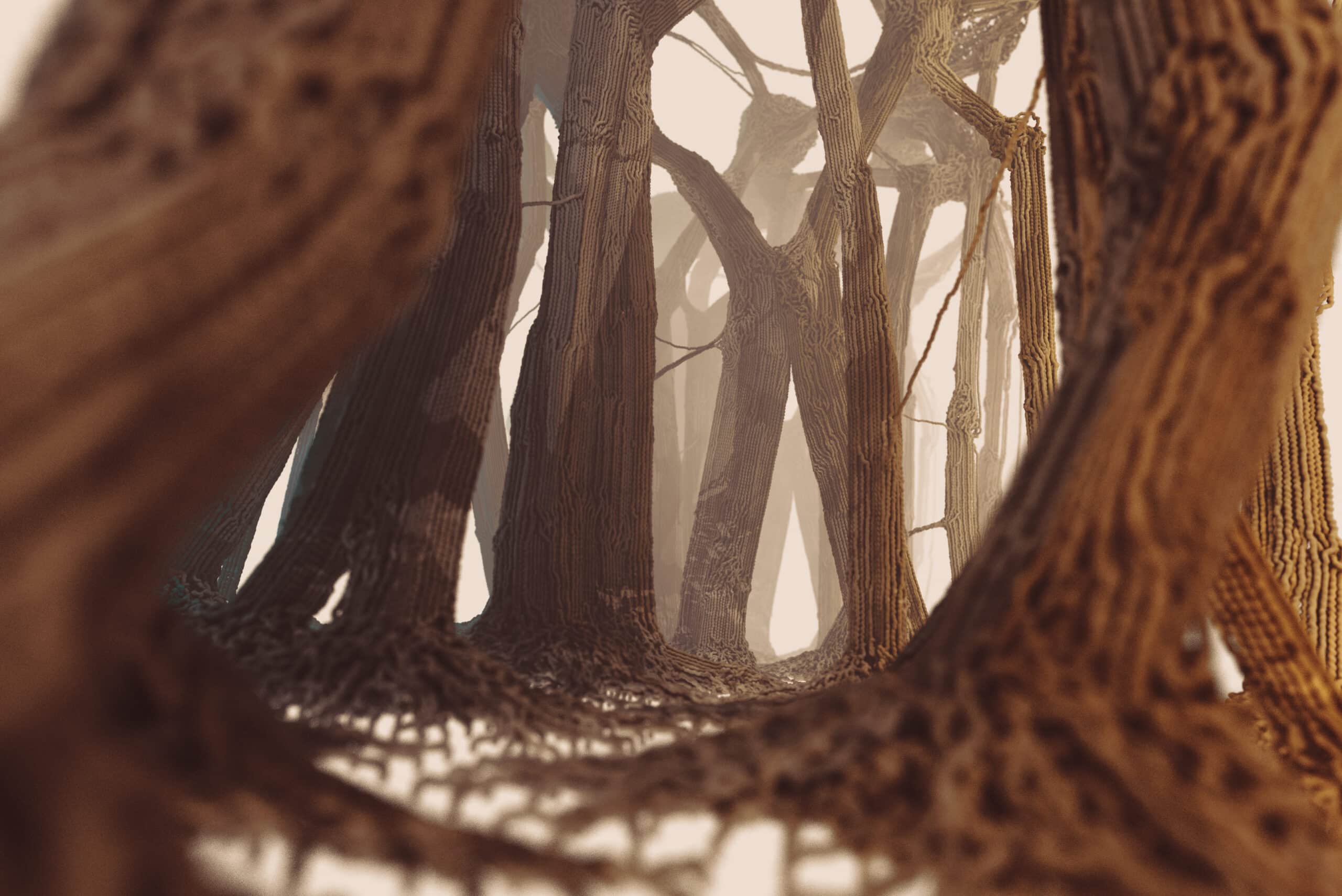
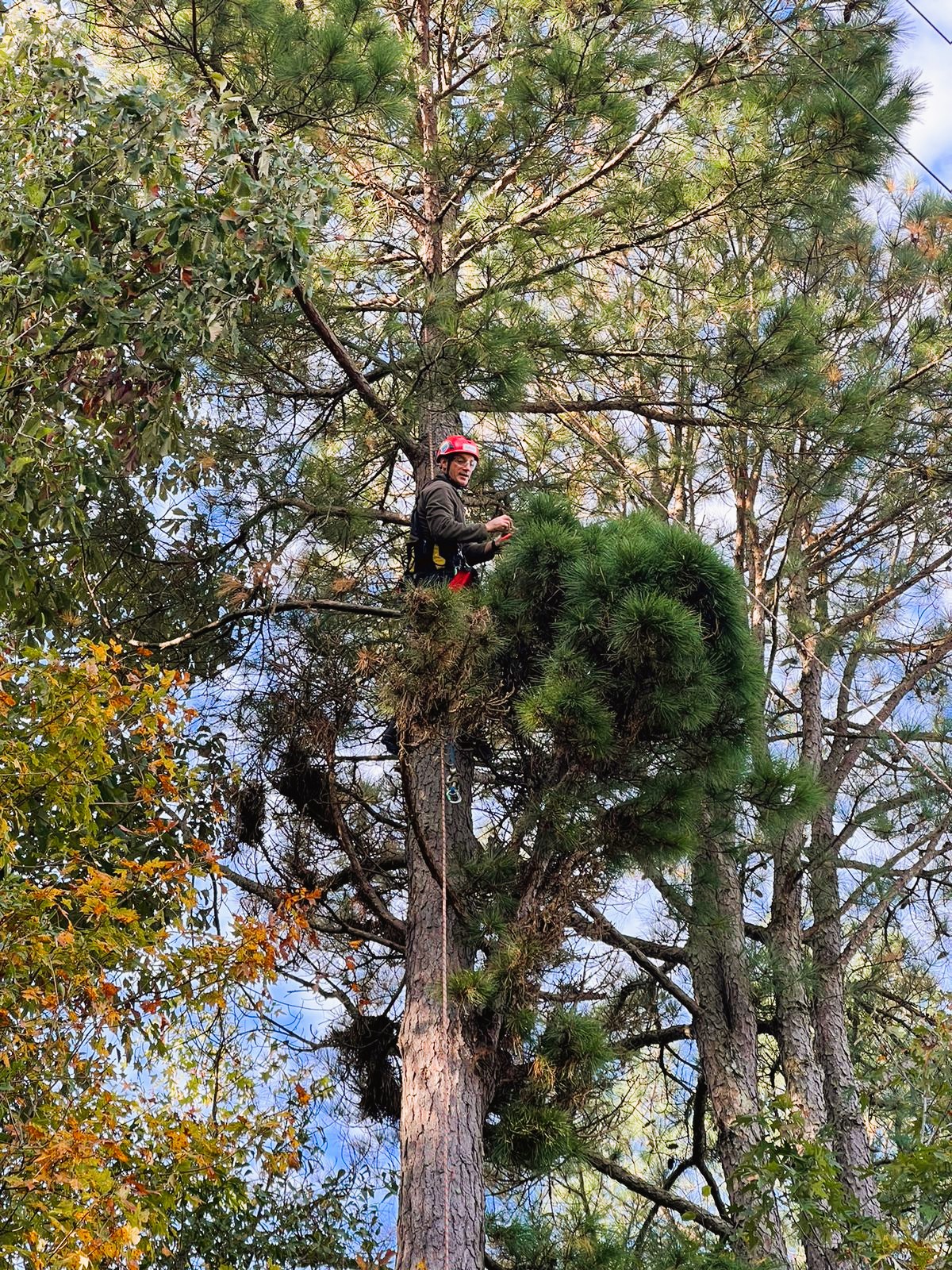
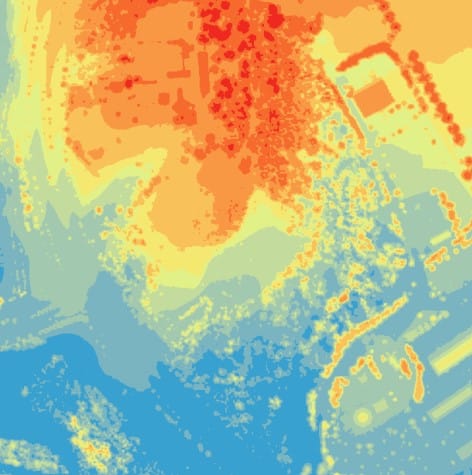
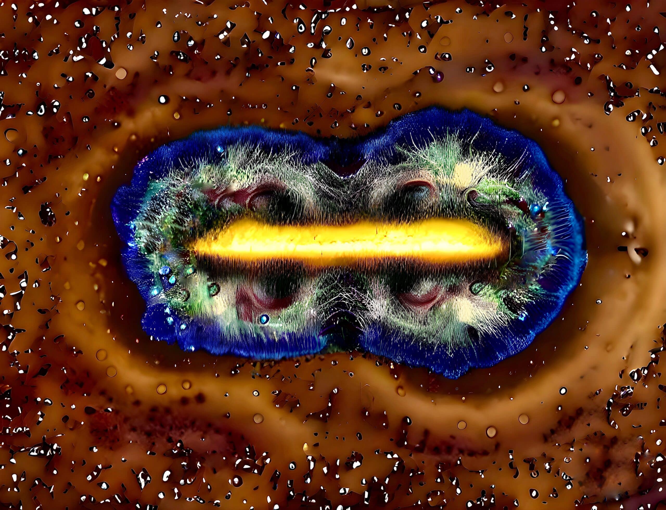
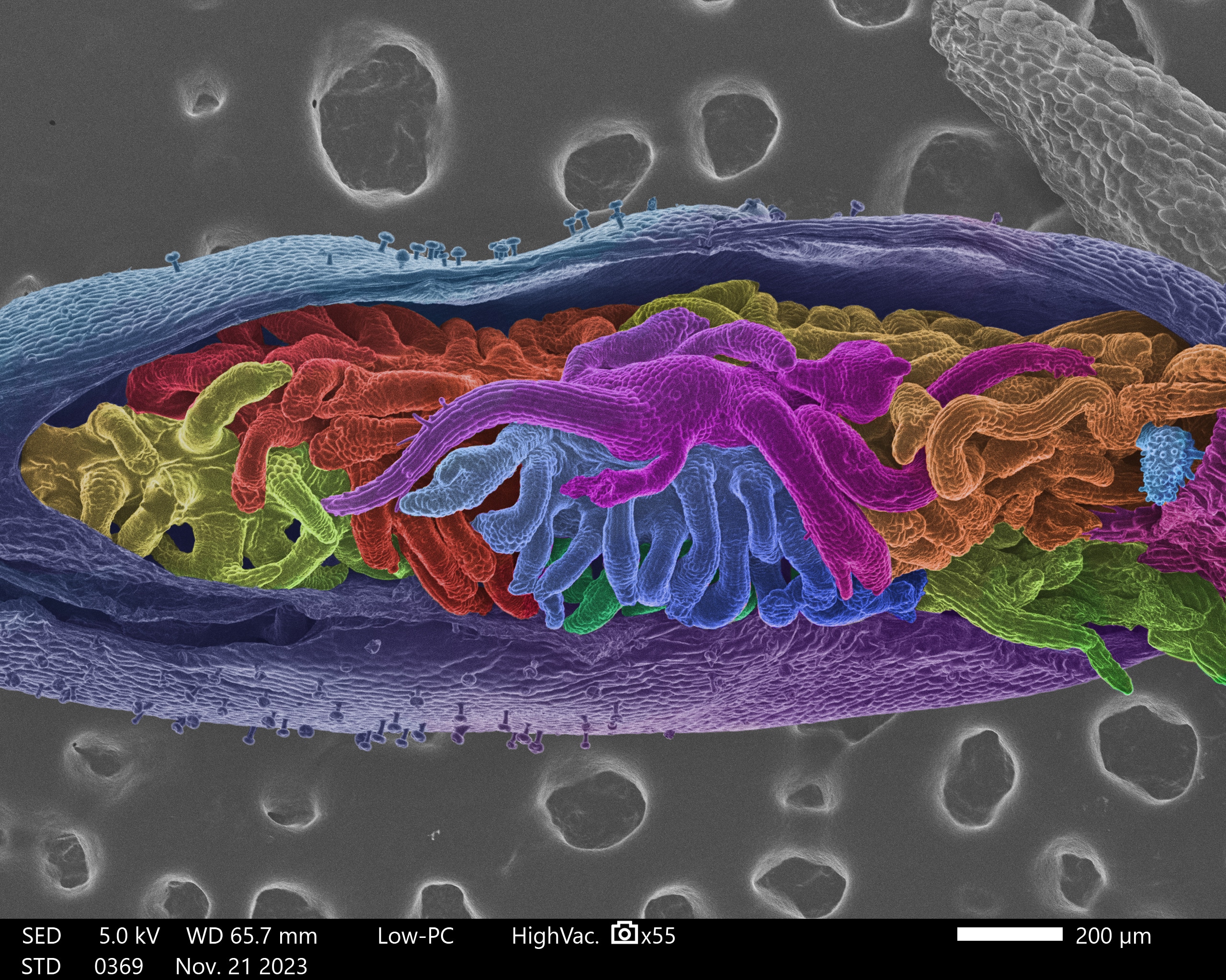
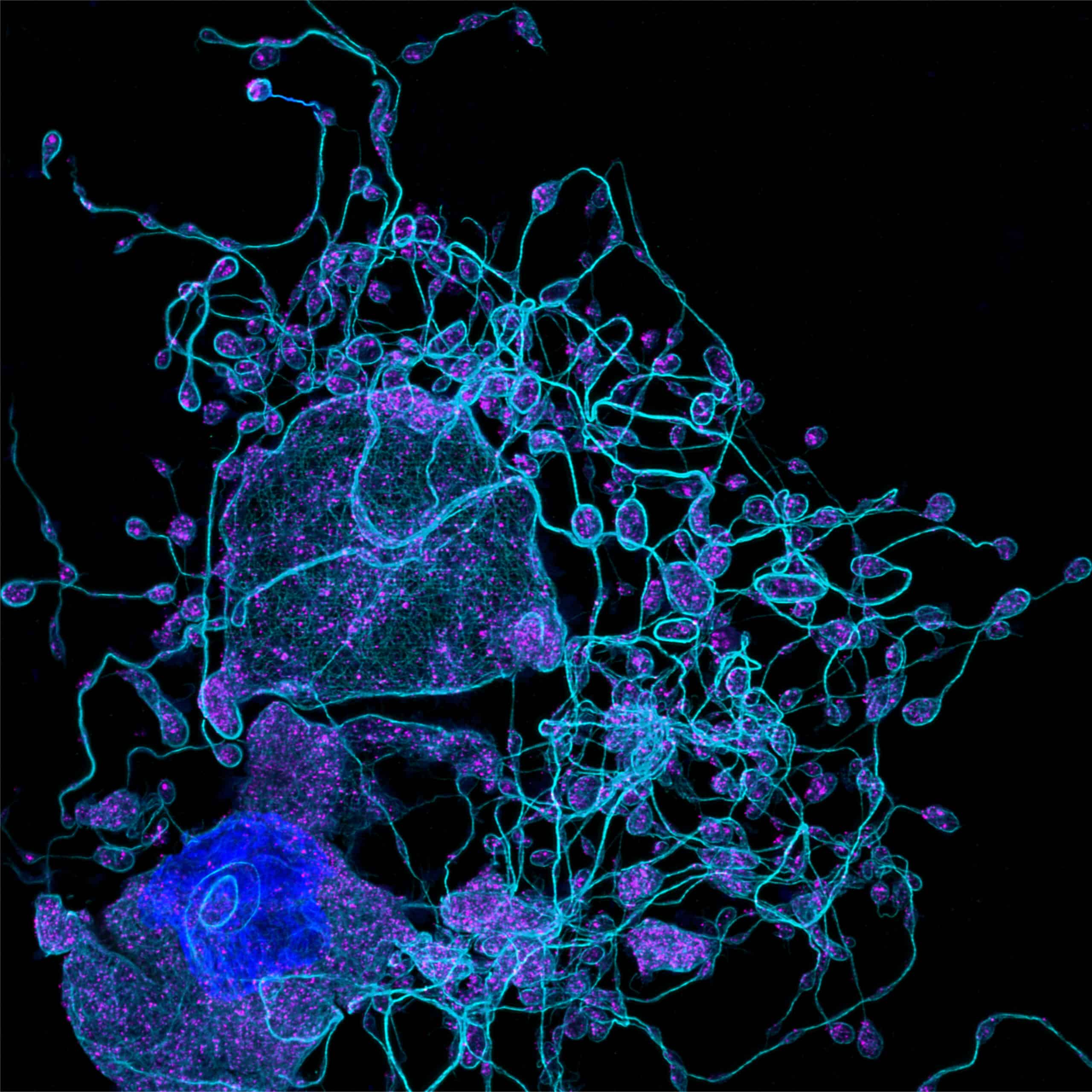
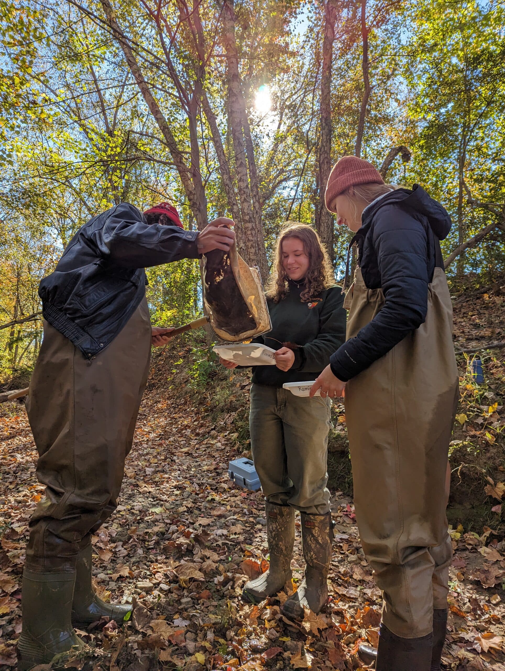
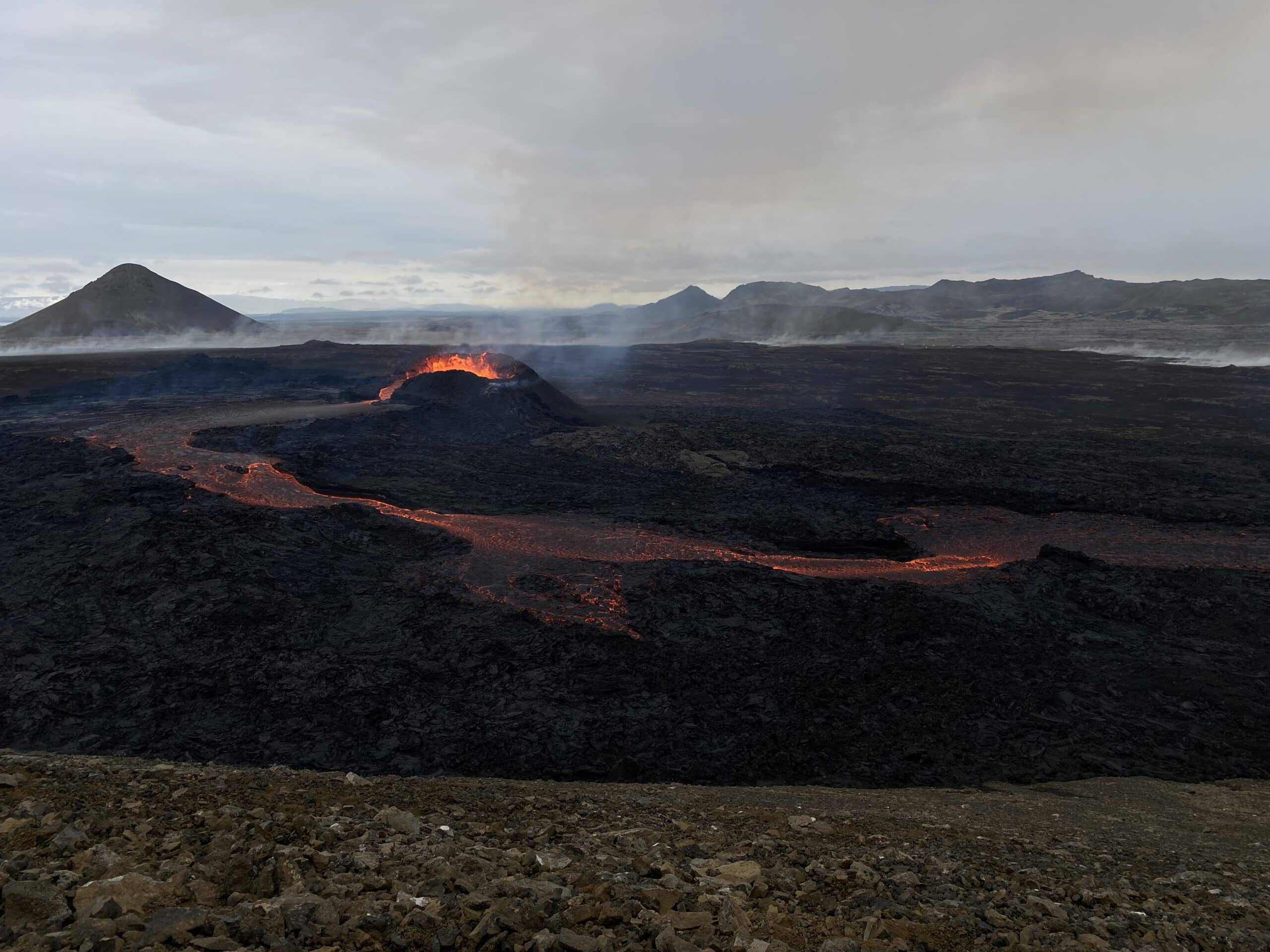
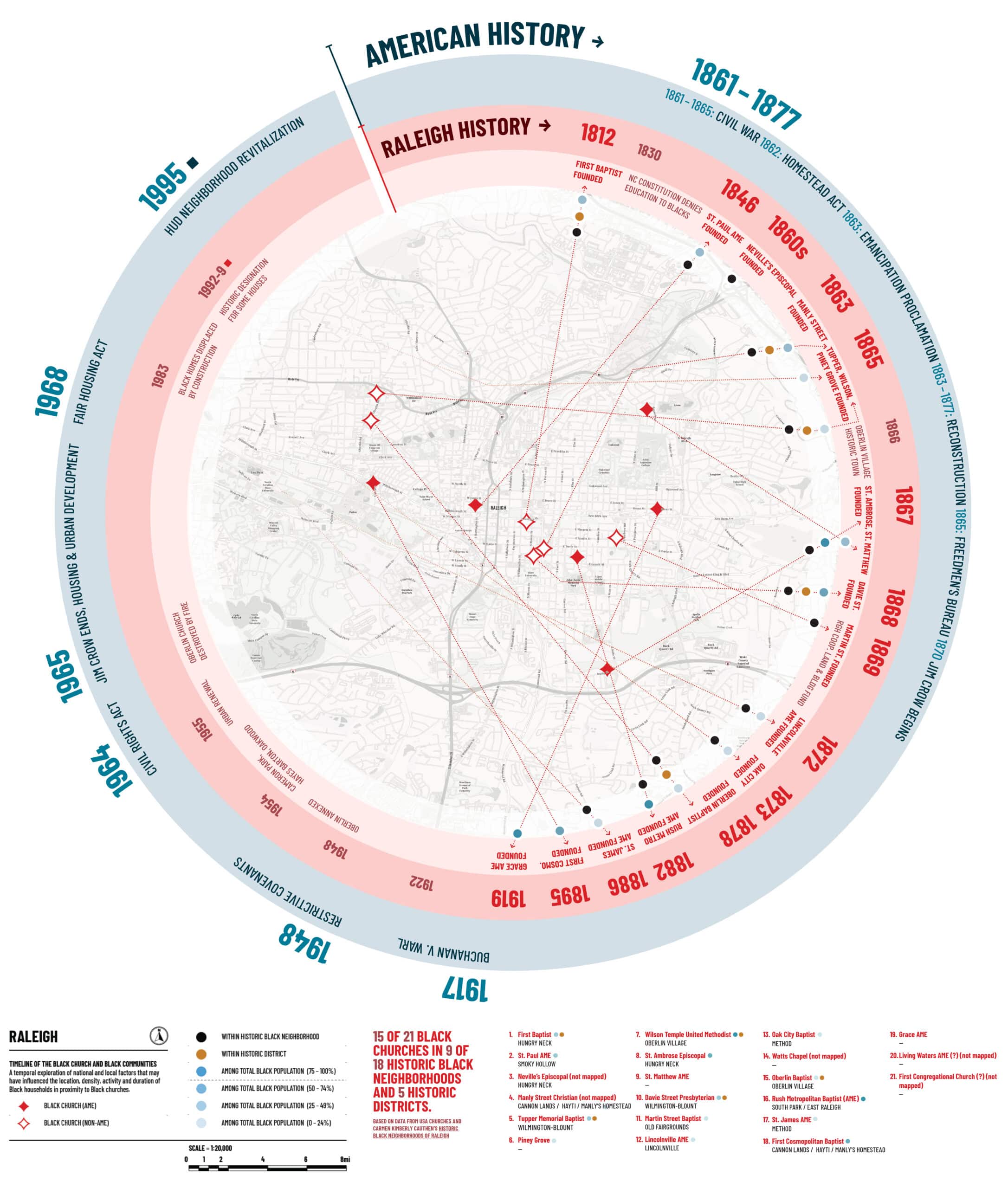
Photography: First place, Graduate Students and Postdocs: Anukram Adhikary for “Frontiers of feminine fortitude.”Microscopy: First place, Undergraduate Students: Zachary Benfield for “Mature Streblospio benedicti headgear.”Photography: First place, Undergraduate Students: Nicolás Galvez for “Cockroach preparing to jump.”Graphics and Data Visualization: First place, Graduate Students and Postdocs: Sergei Rigin for “Molecular forest.”Photography: Second place, Faculty and Staff: Nasir Shalizi for “Needle and cone collection from a witch’s broom 60′ above on a loblolly pine tree.”Graphics and Data Visualization: Honorable mention, Graduate Students and Postdocs: Daoru Wang for “Sakyamuni Pagoda of Fogong Temple.”Photography: Second place, Undergraduate Students: Emily Boldor for “Seismic design structure.”Graphics and Data Visualization: Second place, Graduate Students and Postdocs: Skylar Penney for “Spline image of NC State’s campus.”Microscopy: First place, Graduate Students and Postdocs: Abhirup Basu for “Corrosion’s supernova: unveiling destructive power in coatings.”Microscopy: Second place, Undergraduate Students: Lia Hunt for “Medusa mutation in M. guttatus seed pod.”Microscopy: Second place, Faculty and Staff: Nathan Asquith for “Megasplosion.”Photography: First place, Faculty and Staff: Erin McKenney for “Undergraduate ecologists in the field.”Photography: Honorable mention, Graduate Students and Postdocs: Micki Recchuiti for “Eruption in Iceland: new land by the hour.”Graphics and Data Visualization: Honorable mention, Graduate Students and Postdocs: Ariana Farquharson for “Black churches as community anchors: Raleigh’s Black churches, 1830-present.”
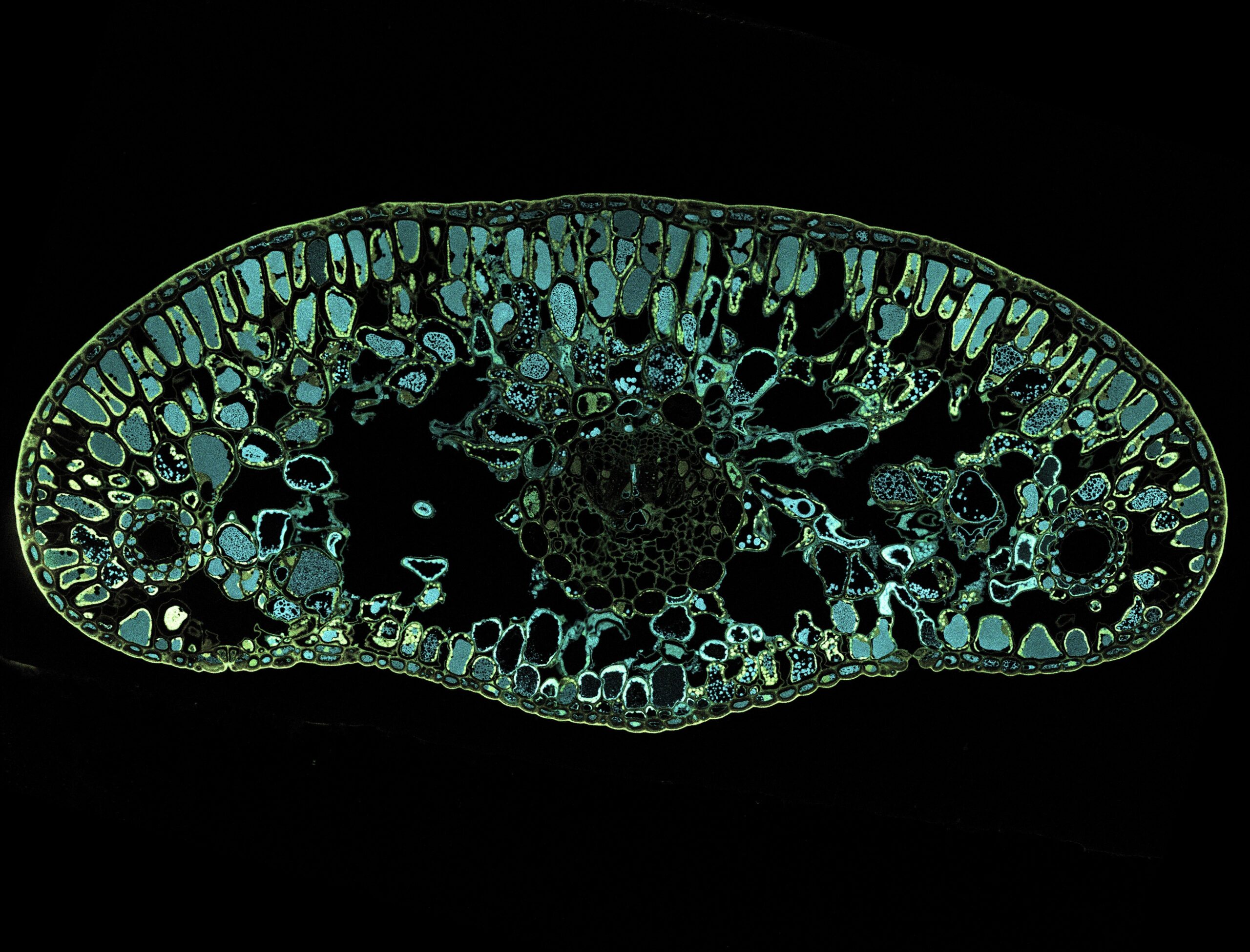
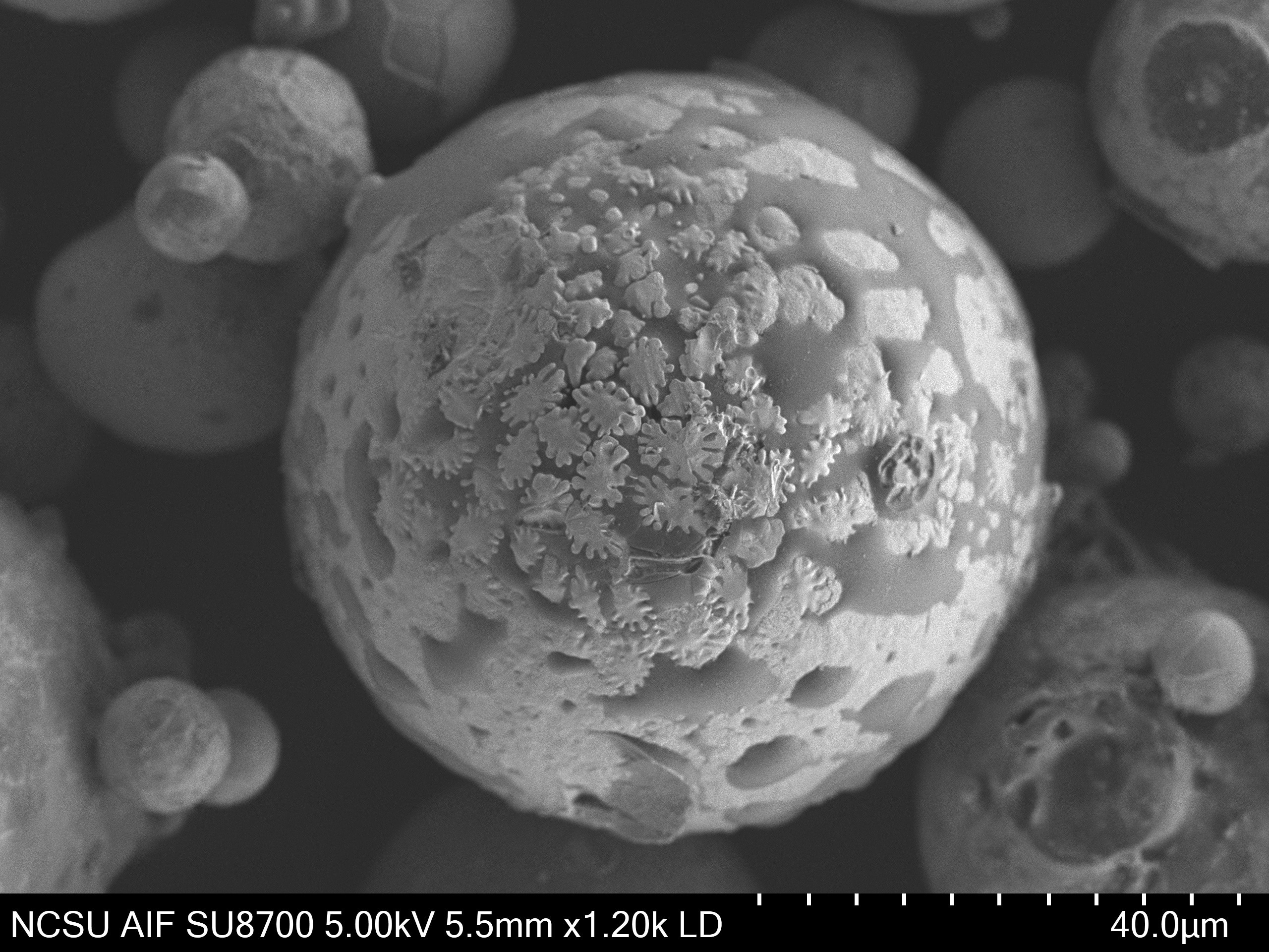
Microscopy: Honorable mention, Graduate Students and Postdocs: Sai Karthik Gade for “Cross section of a Fraser fir needle showing an abundance of polyphenolic cells (blue) induced by phytohormone methyl jasmonate.”Microscopy: Second place, Graduate Students and Postdocs: Victoria Himelstein for “In my own world: dendrite nucleation visible on the outside of a Mo-Si-B powder particle.”Microscopy: First place, Faculty and Staff: Scott LaGreca for “Erysiphe sp. nov. chasmothecium.”
Related
Results Magazine
Your source for the latest research and innovation coming out of NC State.