Nothing Fishy About This Surgery
Cataracts aren’t just a problem for the AARP set. They can affect all kinds of animals, including fish. Veterinary surgeons from North Carolina State University have successfully removed a cataract from a sunfish’s eye, utilizing the same surgical techniques that your local ophthalmologist would use on your grandparents.
The procedure offers a new way for both pet owners and museum curators to safeguard the health of their fish. Let’s say you work at a museum with a collection of fish that you keep for display and educational purposes. Now let’s say that one of your fish has developed a cataract. Impaired vision can affect an animal’s ability to feed, and lead to declining health or even death. Aside from the obvious reasons for keeping animals from needless suffering, sometimes these fish aren’t easily replaceable – they represent significant investments in terms of both money and time.
Greg Lewbart and Brian Gilger are professors of clinical sciences at NC State. Lewbart specializes in aquatic animals – fish, turtles, some reptiles – and Gilger is an expert on eyes. When they came across a five-year-old sunfish named River at the Museum of Life + Science in Durham, N.C. that had a cataract on just one eye, Lewbart decided that River was an excellent candidate for surgery.
“We’d done cataract surgery on reptiles, including sea turtles, and theorized that the technique would work on fish as well,” Lewbart says. “But we needed a fish with just one cataract, so we could be sure that it was due to an injury or cause aside from systemic disease. Often when you see double cataracts in fish, they’re caused by infections or nutritional deficiency, and cataract surgery in those cases wouldn’t solve the problem.”
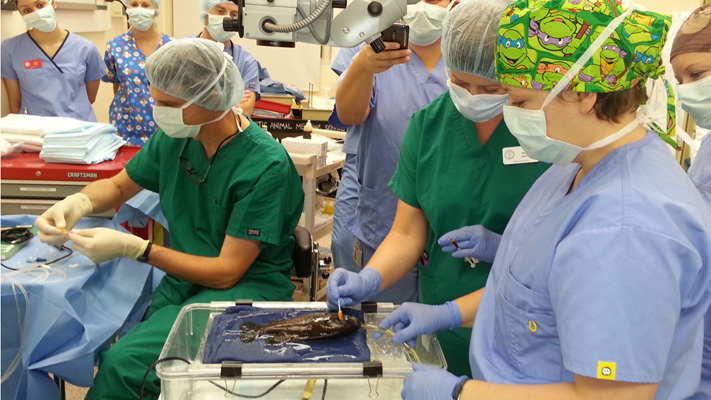
So River the sunfish underwent cataract surgery, which necessitated a very special operating theatre.
The Fish Anesthesia Delivery System, or FADS, is a specially designed operating “table” that gives surgeons access to the fish without taking the animal completely out of its element. Lewbart and fellow NC State colleague Craig Harms developed FADS in the 1990s. Picture a fountain, where water travels up through a tube and then sprays back out. That’s the general idea – the table is like a shallow aquarium with a submersible pump. The fish lays with one side out of the water, covered with a special drape that preserves moisture. An anesthetic is dissolved into the water, which is pumped via a tube through the fish’s gills, so the fish has oxygen and is sedated, and the surgeon can operate.
For the actual surgery, Gilger used the same technique that human ophthalmologists do. It’s called phaecoelmusification and involves “shattering” the cataract with high frequency sound waves and then vacuuming the pieces out of the lens. The only difference in River’s case was that the fish cataract was tougher than a human’s, and so Gilger had to make a small incision to remove some of it. But aside from that one hiccup, the surgery went well and River’s recovery was smooth.
Postdoctoral veterinary intern Dr. Laura Adamovicz also assisted with and co-authored a paper describing the procedure, which appears in the Journal of Fish Diseases.
Lewbart was pleased with the outcome. “People have been doing surgery on fish for a long time, but we have the ability and technology now to bring these surgeries up to a medical standard of care, and to offer them not just to museums but pet owners as well.”
- Categories: