Persistent Photoconductivity Used to Stimulate Neurotypic Cells

For Immediate Release
Researchers have, for the first time, used a material’s persistent photoconductivity to stimulate neurotype cells. The technique, which is relatively simple, should facilitate future research on using charge to influence cellular behavior.
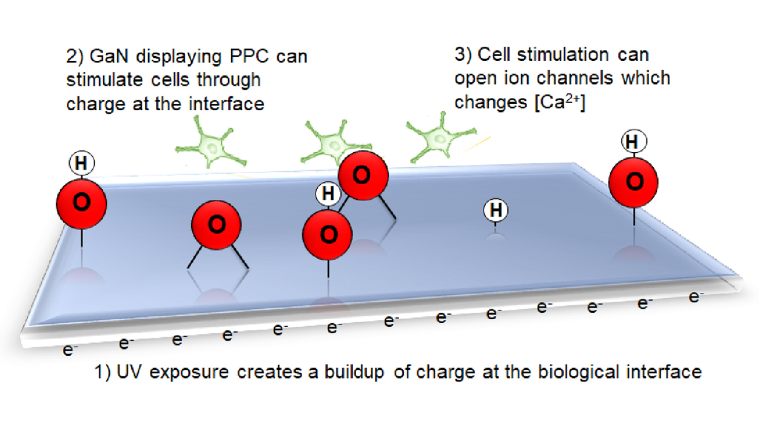
A material that demonstrates persistent photoconductivity gains a negative charge on its surface when exposed to the right wavelength of light, and retains that charge even after the light is removed. The correct wavelength of light, and how long the material retains its charge, varies from material to material.
Researchers have known for years that electric charge can stimulate a cell, but existing technologies for conducting related experiments are often invasive or require specialized equipment. They can also be extremely time consuming.
“We wanted to take advantage of the electronic properties of the semiconductor gallium nitride, which is biocompatible, making it a true bioelectronic interface,” says Patrick Snyder, a Ph.D. student at North Carolina State University and lead author of a paper on the work. “The result is a faster, noninvasive way to stimulate cells that doesn’t require specialized equipment.”
Specifically, the researchers exposed a gallium nitride substrate to ultraviolet (UV) light, creating a negative charge on its surface. As soon as the UV light was removed, researchers poured a solution containing PC12 neurotypic cells into the container with the substrate. The researchers then introduced a dye that allowed them to measure calcium levels in the PC12 cells.
What the researchers found was that PC12 cells were stimulated when they came into contact with the charged gallium nitride substrate, as evidenced by increased calcium ion levels within the cells, when compared to a control group that did not come into contact with a charged substrate.
This is evidence of altered behavior because ions are important in neurotypic cell activity. For example, calcium ions play key roles in neuronal signaling.
“In addition to advancing our fundamental understanding of what this material is capable of, we’re optimistic that it can facilitate work by many labs interested in advancing research on cellular behavior,” says Albena Ivanisevic, a professor of materials science and engineering at NC State and corresponding author of the paper.
The paper, “Non-Invasive Stimulation of Neurotypic Cells Using Persistent Photoconductivity of Gallium Nitride,” is published in the open-access journal ACS Omega. The paper was co-authored by Ramon Collazo, an assistant professor of materials science and engineering at NC State; Pramod Reddy, a postdoctoral researcher at NC State and employee of Adroit Materials; Ronny Kirste of Adroit Materials; and Dennis LaJeunesse, an associate professor of nanoscience at the University of North Carolina-Greensboro.
The work was done with support from the U.S. Army Research Office, under grant W911NF-15-1-0375; and the National Science Foundation, under grants DMR-1312582 and ECCS-1653383.
-shipman-
Note to Editors: The study abstract follows.
“Non-Invasive Stimulation of Neurotypic Cells Using Persistent Photoconductivity of Gallium Nitride”
Authors: Patrick J. Snyder, Ramon Collazo and Albena Ivanisevic, North Carolina State University; Pramod Reddy, North Carolina State University and Adroit Materials; Ronny Kirste, Adroit Materials; Dennis R. LaJeunesse, University of North Carolina-Greensboro
Published: Jan. 19, ACS Omega
DOI: 10.1021/acsomega.7b01894
Abstract: The persistent photoconductivity (PPC) of n-type Ga-polar GaN was used to stimulate PC12 cells non-invasively. Analysis of the III-V semiconductor material by atomic force microscopy, kelvin probe force microscopy, photoconductivity and X-ray photoelectron spectroscopy quantified bulk and surface charge, as well as chemical composition before and after exposure to UV light and cell culture media. The semiconductor surface was made photoconductive by illumination with UV light and experienced PPC which was utilized to stimulate PC12 cells in vitro. Stimulation was confirmed by measuring the changes in intracellular calcium concentration. Control experiments with gallium salt verified stimulation of the neurotypic cells. Inductively coupled plasma mass spectrometry data confirmed the lack of gallium leaching and toxic effects during the stimulation.
- Categories:


