Envisioning Research
Take a look at some of the breathtaking winning images from the 2018 photo and video competition.
Envisioning Research is an annual photo and video competition for research faculty, staff, graduate students and postdocs to celebrate the beautiful images that come from research at NC State.
Visit the contest website for more information, to submit an entry for this year, and to see the full winner galleries.
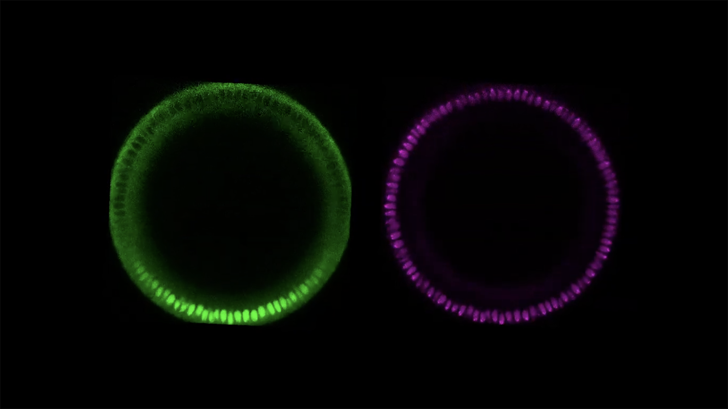
Hadel Al Asafen, Graduate student in the Department of Chemical and Biomolecular Engineering
Live imaging of a 2-hour old fruit fly embryo, imaged frontally using confocal microscopy. The magenta color channel is for the nuclei and the green color channel is for a protein called Dorsal (Dl) that was tagged with GFP. Fruit flies have no cellular membranes separating the nuclei. After nine nuclear divisions, the nuclei migrate to the periphery of the embryo, where they remain dividing until the formation of the ventral furrow and gastrulation (loss of embryonic symmetry). Dorsal (Dl), one of three homologs to mammalian NF-κB, acts as a morphogen to pattern the dorsal-ventral (DV) axis. Dl is initially distributed uniformly along the DV axis. After the 9th nuclear division cycle, when the nuclei migrate to the periphery of the syncytial embryo, Dl begins to accumulate in ventral nuclei, and is excluded from dorsal nuclei. The video clearly shows the formation of the ventral furrow on the ventral side of the embryo, where Dl accumulates. The sides of the furrow will then be brought together into the interior of the embryo to give rise to mesoderm.
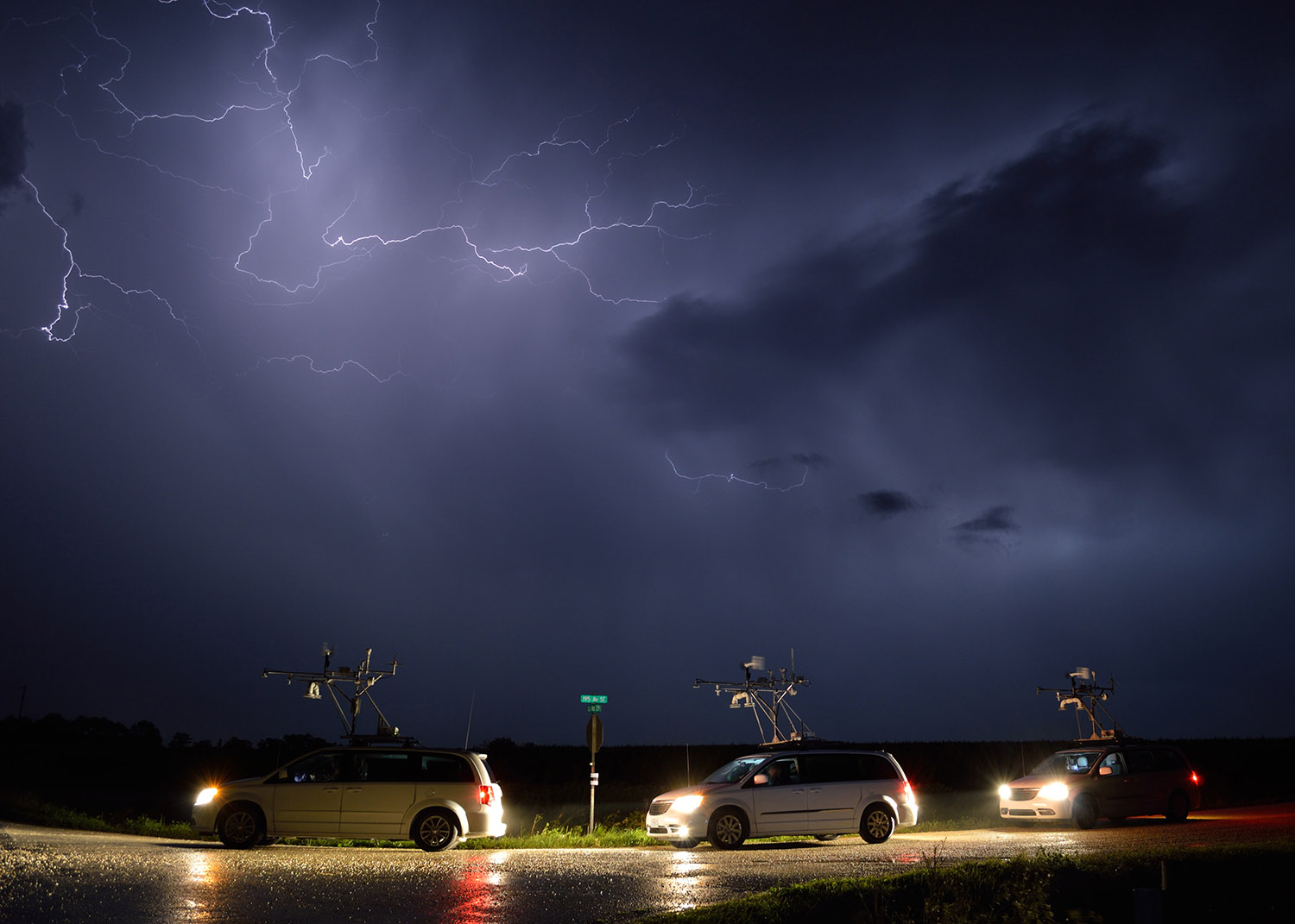
Andy Wade, Ph.D. Student in the Department of Marine, Earth and Atmospheric Sciences
Graduate students from NC State, the University of Oklahoma and Colorado State University collect data on a line of passing storms in southeastern Minnesota. The Plains Elevated Convection at Night experiment sent researchers from numerous universities and laboratories across the central U.S. in search of nocturnal thunderstorms.
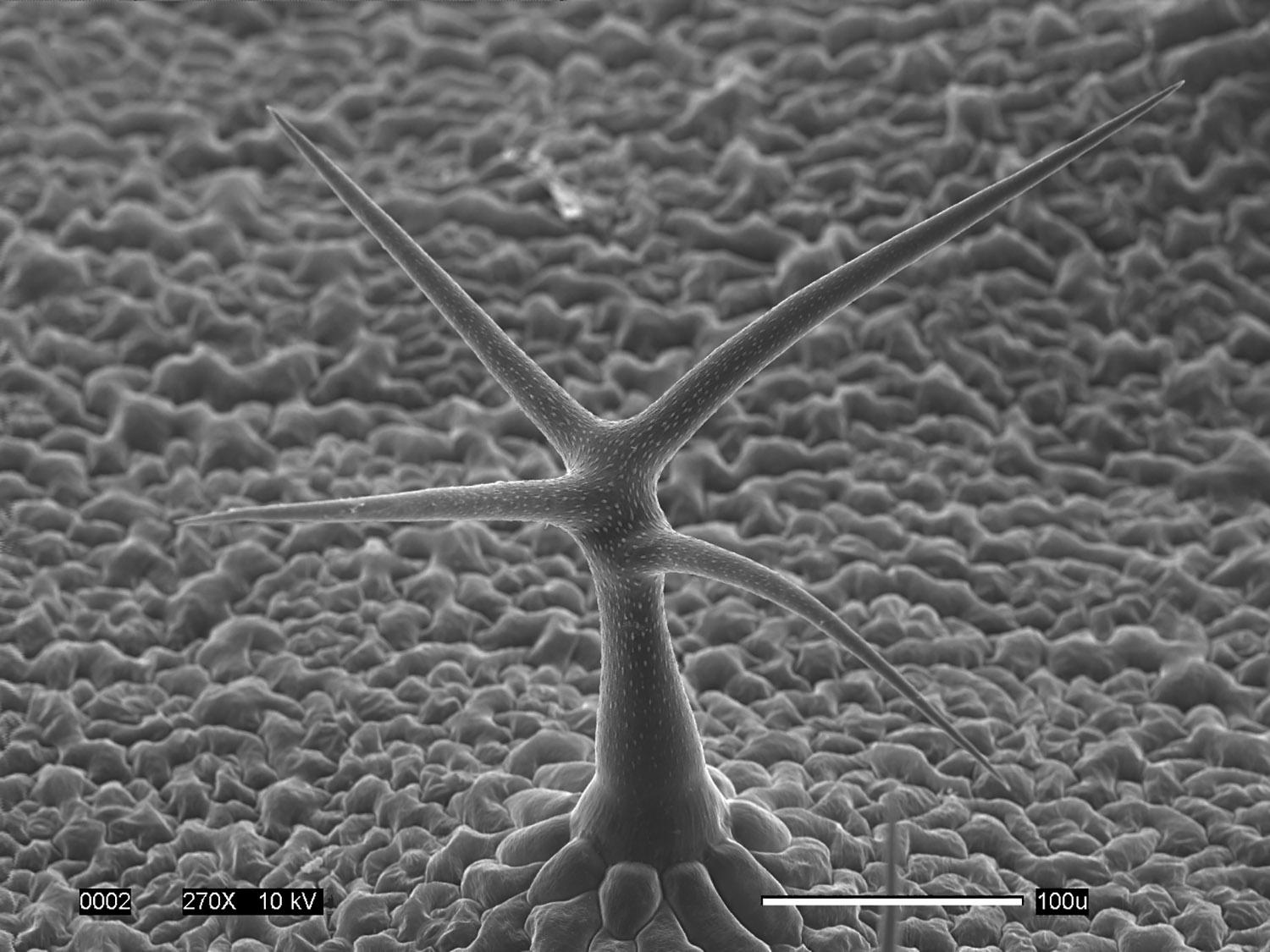
Eric Land, Ph.D. Student in the Department of Plant and Microbial Biology
Arabidopsis Trichome
This scanning electron micrograph captures the impressive three-dimensional ultrastructure of an Arabidopsis “leaf hair” that looks like a tree growing in a meadow. Trichomes like this one are single-cell appendages growing out of the leaf surface.
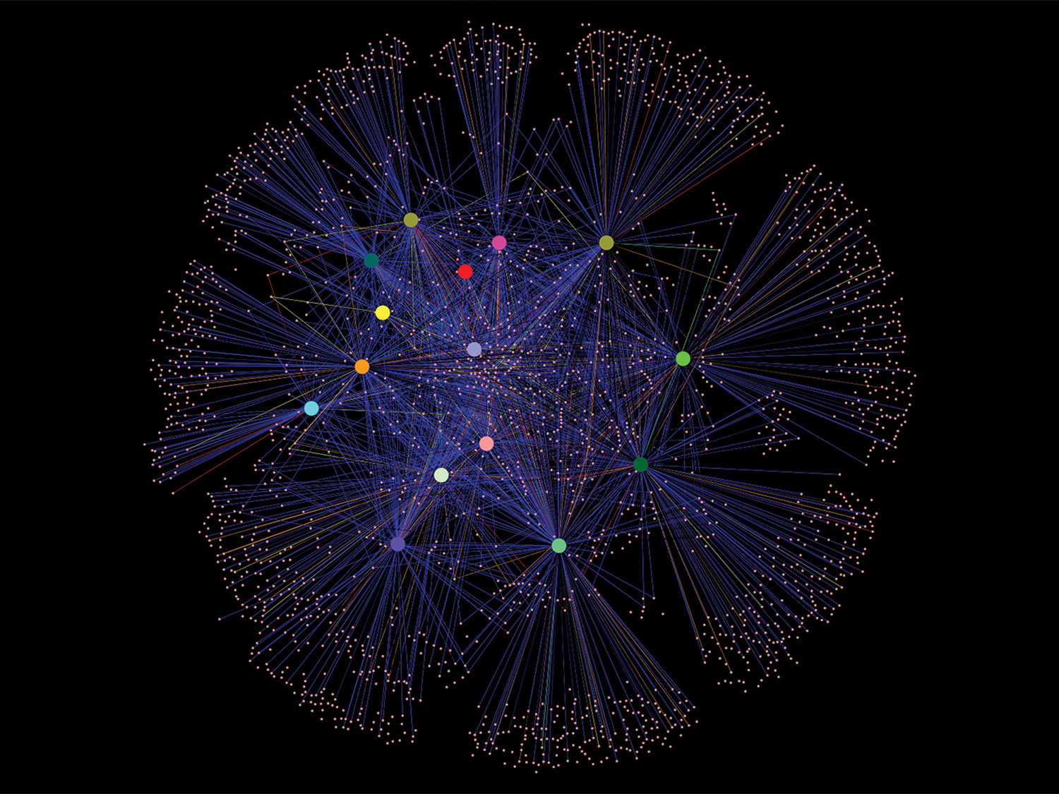
Michael Fisher, Ph.D. Student in the Department of Entomology and Plant Pathology.
This illustration depicts an Operational Taxonomic Unit (OTU) network that shows the diversity of bacteria among bed bug populations around North Carolina. Each colored dot in the center represents a different sample location around the state. The sand-colored dots on the periphery correspond to an individual OTU of bacteria connected by the blue lines, elucidating the relationships. This project examined the gut microbiome of the common bed bug Cimex lectularius from 15 populations around North Carolina.
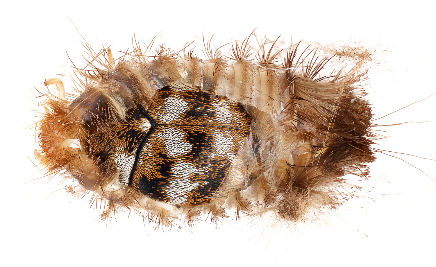
Matt Bertone, Extension Associate in the Department of Entomology and Plant Pathology
Insects and their relatives (called arthropods) are a common — and sometimes abundant — component of the indoor biome. Carpet beetles (Coleoptera: Dermestidae) were found to inhabit 100 percent of the homes sampled in a recent survey of North Carolina homes. This photograph shows an adult beetle that finished development and is resting in its former larval skin.
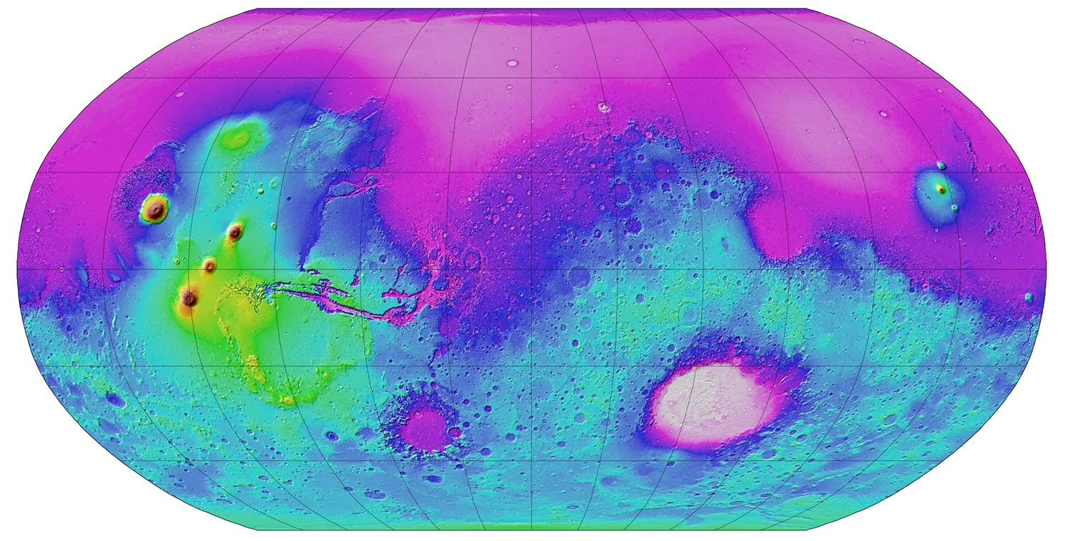
Paul Byrne, Assistant Professor in the Department of Marine, Earth and Atmospheric Sciences
The planet Mars has fascinated humanity for thousands of years. Recent spacecraft missions have returned an unprecedented view of the red planet. Here, topographic data for the entire planet show the vast, low-lying plains to the north, enormous impact basins in the southern hemisphere and, to the west, the largest volcanoes in the solar system—the tallest of which, Olympus Mons, towers 21 kilometers above its surroundings!


