Bone Experts Offer How-To Video For Forensic Professionals
For Immediate Release
Advances in recent years allow forensic practitioners to use bone mineral density to extract more information from human remains – but many forensic experts are unfamiliar with the techniques and technology. Now forensic researchers from North Carolina State University have published a step-by-step methodology in the video journal JOVE, providing forensic professionals with a guide that can help them extract as much information as possible from this emerging tool.
The bone mineral density of a human skeleton can tell forensic practitioners everything from the age of the deceased to whether an individual was malnourished. This information can have multiple uses, such as helping to identify unclaimed remains or determining whether a deceased child was the victim of neglect.
“However, the use of bone mineral density in a forensic context is relatively new, and it is important for practitioners to understand how to use it properly,” says Ann Ross, a professor of biological sciences at NC State and senior author of the journal article. “A video journal allows us to visually demonstrate the process.”
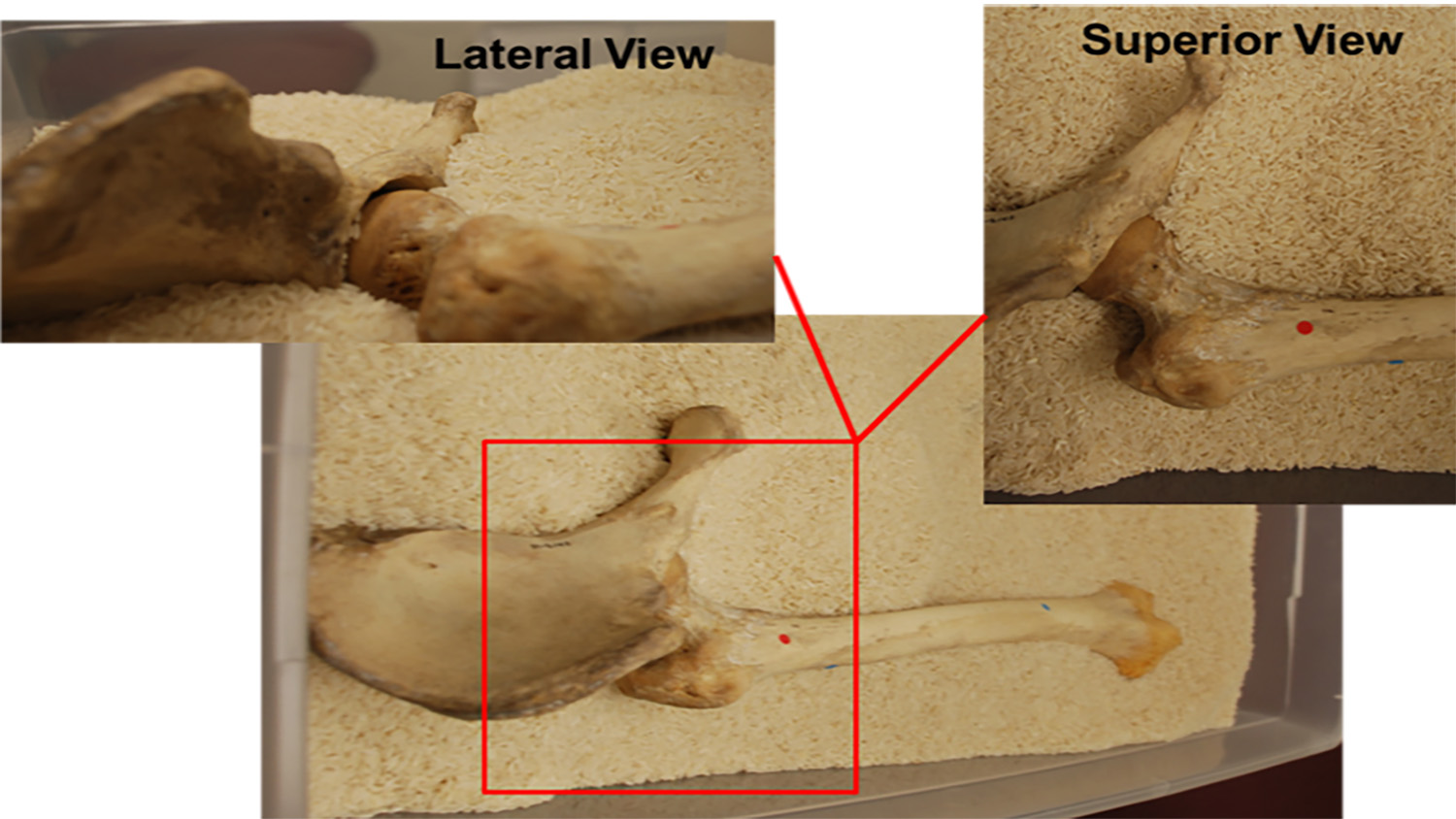
For example, the article explains when collecting bone mineral density may be useful, how to properly scan remains and how to interpret the data.
This is particularly important for collecting and assessing bone mineral density data, both because there are so many variables that can affect that data – and because the data may be used in law enforcement contexts.
“For example, the angle of a bone is important when taking scans, which is much easier to demonstrate via video,” says Amanda Hale, a Ph.D. student in Ross’s lab and first author of the journal article. “And the bone mineral density of remains can be affected by variables such as whether the remains were buried.
“You really need to know when and how you can make measurements that are scientifically robust.”
“The forensic application of bone mineral density is still an active area of research, so knowing how to collect this data properly is likely to become even more important as we learn more about how to properly apply these techniques,” Ross says. “Hopefully our video article will help to ensure that the forensic community is using a consistent suite of methods.”
The paper, “Scanning Skeletal Remains for Bone Mineral Density in Forensic Contexts,” is in press – and available online – in JOVE.
-shipman-
Note to Editors: The study abstract follows.
“Scanning Skeletal Remains for Bone Mineral Density in Forensic Contexts”
Authors: Amanda R. Hale and Ann H. Ross, North Carolina State University
Published: In press, JOVE
Abstract: The purpose of this paper is to introduce a promising, novel method to aid in the assessment of bone quality in forensically relevant skeletal remains. BMD is an important component of bone’s nutritional status and in skeletal remains of both juveniles and adults, and it can provide information about bone quality. For adults remains, it can provide information on pathological conditions or when bone insufficiency may have occurred. In juveniles, it provides a useful metric to elucidate cases of fatal starvation or neglect, which are generally difficult to identify. This paper provides a protocol for the anatomical orientation and analysis of skeletal remains for scanning via dual-energy X-ray absorptiometry (DXA). Three case studies are presented to illustrate when DXA scans can be informative to the forensic practitioner. The first case study presents an individual with observed longitudinal fractures in the weight bearing bones and DXA is used to assess bone insufficiency. BMD is found to be normal suggesting another etiology for the fracture pattern present. The second case study employed DXA to investigate suspected chronic malnutrition. The BMD results are consistent with results from long bone lengths and suggest the juvenile had suffered from chronic malnutrition. The final case study provides an example where fatal starvation in a fourteen-month infant is suspected, which supports autopsy findings of fatal starvation. DXA scans showed low bone mineral density for chronological age and is substantiated by traditional assessments of infant health. However, when dealing with skeletal remains taphonomic alterations should be considered before applying this method.
- Categories:


