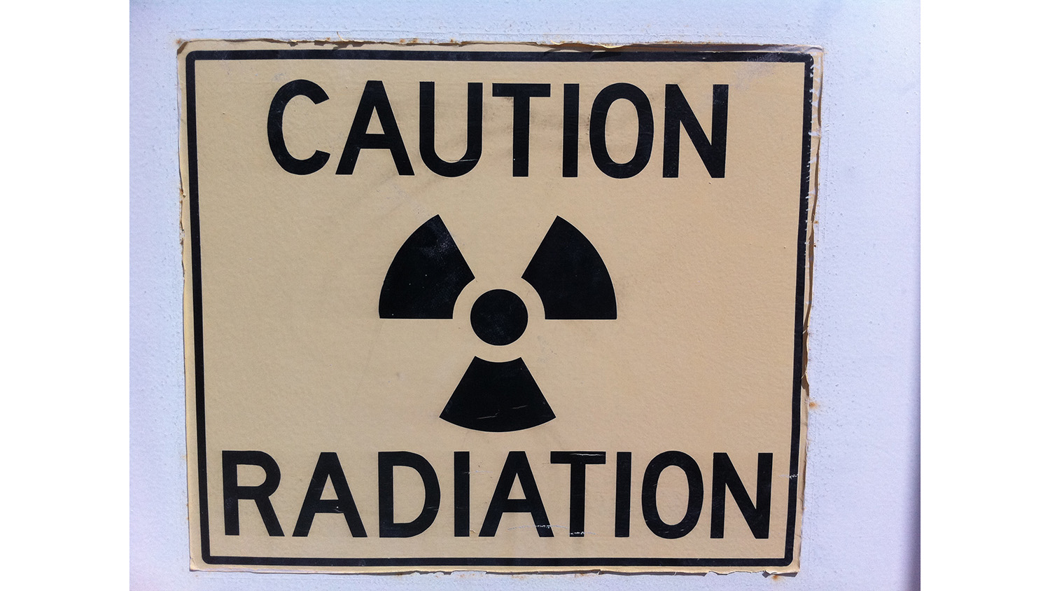
New Technique ‘Sees’ Radioactive Material Even After It’s Gone

For Immediate Release
A new technique allows researchers to characterize nuclear material that was in a location even after the nuclear material has been removed – a finding that has significant implications for nuclear nonproliferation and security applications.
“Basically, we can see nuclear material that is no longer there,” says Robert Hayes, lead author of paper describing the work and an associate professor of nuclear engineering at North Carolina State University. “For example, we could identify and characterize a dirty bomb based on samples taken from a room the bomb was in a year ago.
“This is a valuable tool for emergency responders, nuclear nonproliferation authorities and forensics, because it allows us to get a rough snapshot of the size of a radiation source, where it was located, how radioactive it is, and what type of radioactive material it is,” Hayes says.
The technique takes advantage of the fact that radioactive material changes the arrangement of valence electrons – or outer electrons – in insulator materials, such as brick, porcelain, glass – even hard candy. Basically, radiation displaces electrons at defect sites in the crystalline structure of these materials.
By taking samples of multiple materials in a room, applying conventional radiation dosimetry techniques, and evaluating how the electrons at those defect sites are organized, researchers can determine the presence and strength of any nuclear materials that were in that room.
“If the samples were taken at regular intervals in a grid pattern, the relative radiation dose profile can be used to triangulate where in the room the source was located, in three dimensions,” Hayes says. “It can also provide a very rough idea of the physical size of the source, but that depends on various factors, such as how close the source was to the materials being sampled.”
By taking a core sample of the insulating material, and measuring the radiation dose at various depths in the material, researches can also ascertain what type of radiation source was present. This is possible because different radioactive materials have characteristic distributions of gamma rays, X-rays, etc., and each type of energy penetrates materials with different strength.
“This is not extremely precise, but it does allow us to answer important questions. For example, distinguishing between different kinds of nuclear material such as naturally occurring, medical, industrial, and ‘special’ nuclear materials – the latter being used for nuclear weapons,” Hayes says.
“This is a proof of concept,” Hayes says. “We’re now focused on exploring its detection limitations along with spatial and energy resolution, and how to make use of this approach moving forward.
“But this is a big deal for nuclear nonproliferation efforts, because it means you can’t handle nuclear material in secret anymore,” Hayes adds. “It means the world is now densely blanketed by low-resolution integrating gamma-ray spectrometers, so we can always go back and measure what was present. There’s no hiding.”
The paper, “Retrospective imaging and characterization of nuclear material,” is published in the journal Health Physics. The paper was co-authored by Sergey Sholom of Oklahoma State University. The work was done with support from the Nuclear Regulatory Commission, under grant number NRC-HQ-84-14-G-0059, and Oak Ridge National Laboratory in coordination with the Office of Defense Nuclear Nonproliferation R&D of the National Nuclear Security Administration sponsored Consortium for Nonproliferation Enabling Capabilities.
-shipman-
Note to Editors: The study abstract follows.
“Retrospective imaging and characterization of nuclear material”
Authors: Robert B. Hayes, North Carolina State University; Sergey Sholom, Oklahoma State University
Published: July 3, Health Physics
DOI: 10.1097/HP.0000000000000680
Abstract: Modern techniques for detection of covert nuclear material requires some combination of real time measurement and or sampling of the material. More common is real time measurement of the ionizing emission caused by radioactive decay or through the materials measured response to external interrogation radiation. You can expose the suspect material with various radiation types including high energy photons such as X-rays or with larger particles such as neutrons and muons to obtain images or measure nuclear reactions induced in the material. Stand-off detection using imaging modalities similar to those in the medical field can be accomplished or simple collimated detectors can be used to localize radioactive materials. In all such cases, the common feature is that some or all of the nuclear materials have to be present for the measurement which makes sense as one might ask, “How can you measure something that is not there?” The current work and results show how to do exactly that, how to characterize nuclear materials after they have been removed from an area leaving no chemical trace. This new approach is demonstrated to be fully capable of providing both previous source spatial distribution and emission energy grouping. The technique uses magnetic resonance for organic insulators and/or luminescence techniques on ubiquitous refractory materials similar in theory to the way the nuclear industry carries out worker personnel dosimetry. Spatial information is obtained by acquiring gridded samples for dosimetric measurements while energy information comes through dose depth profile results which are functions of the incident radiation energies.
- Categories:


